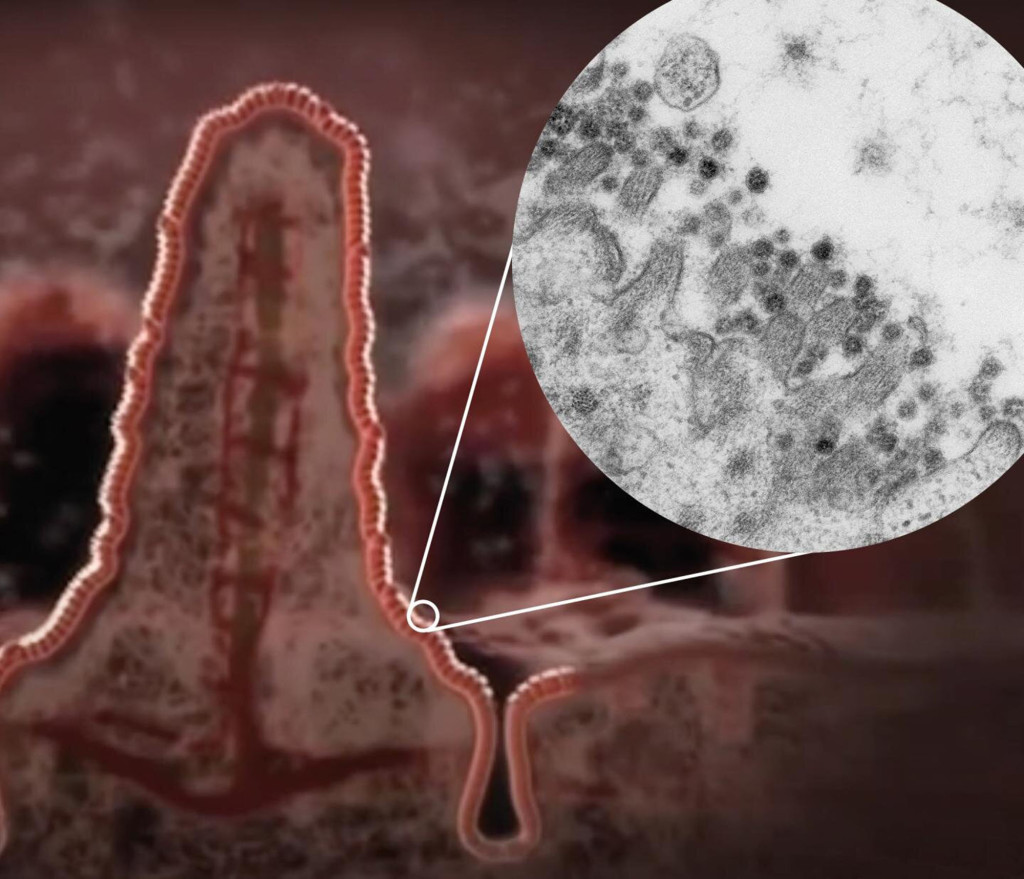
The first detailed, glowing images have emerged of the novel coronavirus – showing the deadly pathogen multiplying in the gut, according to a report.
A British team at the University of Dundee’s School of Life Sciences released some of the sharpest photos yet recorded of the bug, which has killed over 247,000 people around the world, The Sun reported.
The images from a powerful microscope show the formation of Sars-CoV-2 particles in a tissue model of a person’s gut and how the virus assembles and leaves intestinal cells.
The scientists collaborated with experts at the Hubrecht Institute in Utrecht, the Erasmus MC University Medical Centre in Rotterdam and Maastricht University in the Netherlands to isolate the highly detailed images.
Led by Dundee’s Professor Jason Swedlow, the team discovered that the virus can infect the cells of the intestine and multiply there.
The researchers also successfully grew the virus in test tubes and monitored the response of the cells, with findings that could explain why about a third of patients experience gastrointestinal symptoms such as diarrhea and how the virus can often be detected in stool samples.
“We’re excited to publish these important new datasets in IDR, where they can be seen by researchers around the world, who can also scan the images and view the Sars-CoV-2 virus up close on their computer,” Swedlow said, referring to the Image Data Resource.
“We have included annotations from the authors so anyone who reads the paper from the research teams in the Netherlands can easily see what the authors published, but also can examine other parts of the data and maybe make their own discoveries,” the cell biologist said.
“This kind of sharing of data has never been more important than in our current situation where we urgently need to work together around the world to find out more about this disease and ultimately be able to treat or control it,” Swedlow added.


